
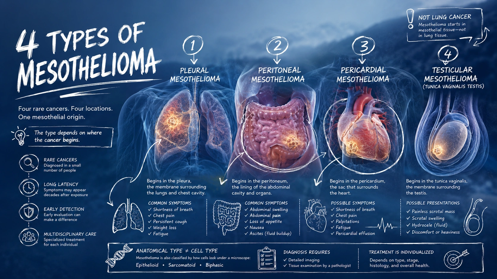
Mesothelioma is often described informally as a form of lung cancer, but that description is medically inaccurate. Lung cancer begins in lung tissue. Mesothelioma begins in mesothelial tissue, the thin membrane that lines and protects certain internal organs and body cavities.
The location where the cancer begins determines its type.
The four main anatomical types are pleural, peritoneal, pericardial, and testicular mesothelioma. Pleural mesothelioma, which develops around the lungs, is by far the most common. Peritoneal mesothelioma develops in the abdominal lining, while cancers originating in the pericardium around the heart and the tunica vaginalis around the testis are exceptionally rare.
The differences go beyond anatomy. Each type can produce different symptoms, present different diagnostic challenges, and require a different treatment strategy.
The 4 Types of Mesothelioma at a Glance
The four types of mesothelioma are classified according to where the cancer originates:
- Pleural mesothelioma begins in the pleura, the membrane associated with the lungs and chest cavity.
- Peritoneal mesothelioma begins in the peritoneum, the membrane lining the abdominal cavity and covering many abdominal organs.
- Pericardial mesothelioma begins in the pericardium, the sac surrounding the heart.
- Testicular mesothelioma, more precisely called mesothelioma of the tunica vaginalis testis, begins in a mesothelial membrane surrounding the testis.
These categories describe the primary site of the cancer. They should not be confused with the histological cell types—epithelioid, sarcomatoid, and biphasic—which describe how the tumor cells appear under a microscope.
Understanding that distinction makes the rest of the disease easier to follow.
1. Pleural Mesothelioma
Pleural mesothelioma is the most common form of malignant mesothelioma. It develops in the pleura, a thin membrane associated with the lungs and the inside of the chest wall.
The pleura consists of two layers. The visceral pleura closely covers the lungs, while the parietal pleura lines the inner surface of the chest wall. A small amount of lubricating fluid between these layers helps them move smoothly against each other as the lungs expand and contract.
When malignant mesothelioma develops in the pleura, tumor growth and inflammation can interfere with this system. Fluid may accumulate in the pleural space, a condition known as pleural effusion. Tumor growth can also produce progressive thickening and restriction around the lung.
Common symptoms can include:
- shortness of breath;
- chest pain;
- persistent cough;
- unexplained weight loss;
- fatigue;
- difficulty taking a deep breath.
These symptoms are not specific to mesothelioma. Many more common diseases can cause similar problems, so symptoms alone cannot establish a diagnosis.
Why Pleural Mesothelioma Is Not Lung Cancer
The distinction between pleural mesothelioma and lung cancer is more than a matter of terminology.
Pleural mesothelioma originates in mesothelial cells of the pleura. Primary lung cancers arise from cells within lung tissue or the airways. The two diseases differ in their biology, staging systems, and treatment strategies.
Asbestos exposure is the major established risk factor for malignant mesothelioma. The disease can emerge decades after exposure, which is one reason diagnoses continue to occur long after the heaviest periods of occupational asbestos use.
How Pleural Mesothelioma Is Diagnosed and Staged
Evaluation may begin with imaging when symptoms or a history of exposure raises concern. CT, PET/CT, MRI, and other tests may be used depending on the clinical situation.
A definitive diagnosis generally requires examination of tumor tissue by a pathologist. The process can be challenging because mesothelioma must be distinguished from metastatic cancers and other conditions involving the pleura.
Among the anatomical forms of mesothelioma, pleural mesothelioma is the only one with a formal widely used staging system. Staging considers the extent of the primary tumor, lymph node involvement, and distant spread.
Treatment depends on disease extent, tumor histology, overall health, and whether the cancer can be treated with a potentially multimodal approach. Depending on the individual case, treatment may involve systemic therapy, surgery in carefully selected patients, radiation therapy, symptom-directed procedures, and palliative care.
2. Peritoneal Mesothelioma
Peritoneal mesothelioma develops in the peritoneum, not in the lungs.
The peritoneum is the serous membrane associated with the abdominal cavity and many abdominal organs. Its parietal layer lines the abdominal wall, while its visceral layer covers many of the organs within the abdomen.
A small amount of fluid normally allows these surfaces to move smoothly. When malignant disease affects the peritoneum, fluid can accumulate in the abdomen. This buildup is called ascites.
Symptoms of peritoneal mesothelioma may include:
- increasing abdominal size or swelling;
- abdominal pain;
- loss of appetite;
- nausea or changes in bowel function;
- unexplained weight loss;
- fatigue;
- ascites.
Because these symptoms overlap with many gastrointestinal, liver, ovarian, and other abdominal conditions, diagnosis can be delayed.
Imaging can identify suspicious abnormalities or fluid accumulation, but tissue examination is generally needed to establish the diagnosis and distinguish mesothelioma from other malignancies involving the peritoneum.
How Peritoneal Mesothelioma Treatment Differs
The treatment pathway for peritoneal mesothelioma can differ substantially from that of pleural disease.
For selected patients, specialist centers may consider cytoreductive surgery combined with hyperthermic intraperitoneal chemotherapy, commonly abbreviated as CRS and HIPEC. Cytoreductive surgery attempts to remove visible tumor deposits, after which heated chemotherapy is circulated within the abdominal cavity.
This approach is not appropriate for every patient. Eligibility depends on factors including disease distribution, tumor biology, whether adequate cytoreduction appears feasible, overall health, and the experience of the treatment center.
Research has reported substantially improved outcomes among carefully selected patients treated with CRS and HIPEC compared with historical outcomes, but the evidence comes largely from observational studies and specialized treatment programs rather than randomized trials. That distinction matters when interpreting survival figures.
3. Pericardial Mesothelioma
Pericardial mesothelioma develops in the pericardium, the membrane surrounding the heart. It is an exceptionally rare cancer, and much of the medical literature consists of case reports, case series, and retrospective database analyses.
The pericardium has two closely related layers with a small amount of lubricating fluid between them. This structure helps reduce friction as the heart moves.
Cancer arising in the pericardium can interfere with normal cardiac function. Depending on the disease pattern, patients may develop pericardial effusion, in which excess fluid accumulates around the heart. Severe fluid accumulation can impair the heart’s ability to fill and pump effectively.
Possible symptoms include:
- shortness of breath;
- chest pain;
- palpitations;
- fatigue;
- cough;
- swelling associated with impaired cardiac function.
These symptoms can resemble much more common heart and lung conditions. That lack of specificity, combined with the rarity of the disease, makes diagnosis particularly difficult.
What Causes Pericardial Mesothelioma?
The relationship between asbestos exposure and pericardial mesothelioma is less straightforward than it is for pleural mesothelioma.
Because the disease is so rare, establishing risk factors through large epidemiological studies is difficult. Research has examined a possible association with asbestos exposure, but the evidence base is much smaller and more uncertain than the evidence for pleural disease.
This is an important place for careful language. It is reasonable to ask about occupational and environmental exposure histories during evaluation, but it is not accurate to assume that every pericardial mesothelioma case was caused by asbestos.
Treatment is also difficult to standardize because prospective clinical evidence is extremely limited. Management may involve surgery, systemic therapy, radiation in selected circumstances, drainage of pericardial fluid, and other supportive interventions. Decisions generally require individualized assessment by a multidisciplinary team.
4. Testicular Mesothelioma
Testicular mesothelioma is commonly used as shorthand for malignant mesothelioma of the tunica vaginalis testis.
The tunica vaginalis is a mesothelial-lined sac associated with the testis. Like other serous membranes, it contains parietal and visceral layers that can move against each other.
Mesothelioma originating at this site is exceptionally rare. Because the total number of documented cases is small, researchers have far less evidence about its causes, natural history, and optimal treatment than they do for pleural mesothelioma.
Possible presentations include:
- a painless scrotal mass;
- scrotal swelling;
- hydrocele, or fluid accumulation around the testis;
- discomfort or a feeling of heaviness;
- an incidental abnormality discovered during surgery for another suspected condition.
The diagnosis may not be apparent before surgery because common conditions such as hydrocele and other scrotal masses can initially produce similar findings.
Is Testicular Mesothelioma Related to Asbestos?
Research has investigated a relationship between asbestos exposure and mesothelioma of the tunica vaginalis testis. Some epidemiological evidence supports an association, but the disease’s extreme rarity makes precise risk estimates difficult.
Not every reported patient has a documented history of asbestos exposure, and exposure information is incomplete in many published cases. Researchers have also investigated other possible contributing factors, including chronic inflammation, but the evidence remains limited.
Another distinction is important: a primary mesothelioma originating in the tunica vaginalis is not the same as a cancer that started elsewhere and later metastasized to structures in the scrotum.
Treatment commonly centers on surgical management when disease is localized, but there is no evidence base comparable to the large clinical trials available for many common cancers. Because recurrence and spread can occur, management and follow-up should involve clinicians experienced with rare genitourinary and mesothelial malignancies.
Anatomical Type and Cell Type Are Different Classifications
A person researching the “types of mesothelioma” will encounter two different classification systems.
The first is anatomical location:
- pleural;
- peritoneal;
- pericardial;
- testicular or tunica vaginalis.
The second is histological type, based on how tumor cells appear under a microscope:
- epithelioid mesothelioma, characterized predominantly by epithelioid tumor cells;
- sarcomatoid mesothelioma, characterized by spindle-shaped malignant cells and generally more aggressive behavior;
- biphasic mesothelioma, containing both epithelioid and sarcomatoid components.
A patient can therefore have, for example, epithelioid pleural mesothelioma or biphasic peritoneal mesothelioma. The anatomical location and histological classification provide different information and can both influence treatment planning and prognosis.
What About “Benign Mesothelioma”?
Older medical terminology can create confusion.
Some tumors historically called “benign mesothelioma” are now classified and understood differently from malignant mesothelioma. For example, solitary fibrous tumor of the pleura was once called localized fibrous mesothelioma, but it is now recognized as a different tumor entity.
This matters because a noncancerous pleural tumor should not automatically be described as a benign form of malignant pleural mesothelioma.
The same caution applies to several rare mesothelial proliferations and tumors of low or uncertain malignant potential. Modern diagnosis depends on pathological classification rather than simply dividing every mesothelial lesion into “benign mesothelioma” and “malignant mesothelioma.”
Why the Type of Mesothelioma Matters
All four anatomical forms originate in mesothelial tissue, but they should not be treated as interchangeable versions of the same clinical problem.
A pleural tumor can restrict the lung and produce pleural effusion. A peritoneal tumor can spread across abdominal surfaces and cause ascites. A pericardial tumor can interfere with the heart through effusion and constriction. A tumor of the tunica vaginalis may first appear as a scrotal mass or hydrocele.
Those differences shape the diagnostic process and treatment options.
They also explain why statistics from one type should not automatically be applied to another. Pleural mesothelioma has a much larger evidence base and a formal staging system. Evidence for pericardial and testicular disease is necessarily based on much smaller patient populations.
For anyone being evaluated for suspected mesothelioma, the most useful questions are therefore more specific than simply, “Is this mesothelioma?” The location of origin, pathological diagnosis, histological subtype, extent of disease, and overall health all help determine what comes next.
Mesothelioma is rare, but its four anatomical forms are not merely different names for the same disease in different places. Each creates its own clinical challenges, and understanding those differences is the first step toward interpreting symptoms, research findings, and treatment information accurately.
- About the Author
- Latest Posts

Salwa Alifah Yusrina is the Editor-in-Chief at Medslat.com. She is a professional Nutritionist with over 7 years of clinical and research experience. She holds a Master’s degree in Nutrition Science from Hasanuddin University. At Medslat.com, she oversees the editorial direction and ensures that all health and nutrition content adheres to the highest standards of accuracy and evidence-based science.